ALEMBIC
GFP-Imaging (Widefield Imaging Setup)
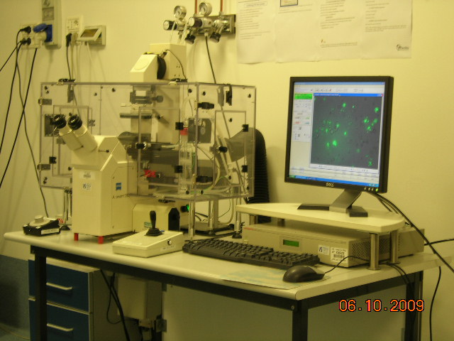
Zeiss Axiovert S100 TV2 with Hamamatsu OrcaII - ER

Objectives
- Plan - NEOFLUAR 5X (NA 0.15) Dry
- A - Plan 10X (NA 0.25) Dry Ph1
- LD Achrostigmat 20X (NA 0.4 3 ) Dry Ph1
- LD AchroPlan 32X (NA 0.4) Dry Ph2
- Plan - APOCHROMAT 63X (NA 1.4 ) Oil DIC
Specifics
- Brightfield (contrast): yes (PH, DIC);
- Fluor: yes;
- Sensor: CCD Camera;
- Max resolution: 1200x1024.
- Z section: yes;
- Time lapse: yes;
- ROI Bleaching: no;
- Other: automated filter-wheels, motorized stage, cage incubator.
Description
Several imaging modes brightfield, phase-contrast, differential-interference contrast (or Nomarski), epi-fluorescence image acquisition with a chilled b&w Hamamatsu CCD camera OrcaII-ER.
Oko-Vision software (from OkoLab) is designed to automatically control all the hardware components of our microscopy workstation and to perform Time-Lapse experiments. This microscope system permits to go from the simple 2D image acquisition to the Multi-dimensional and Multi-channel Live Cell Microscopy.
The Microscope Cage Incubator is designed to maintain all the required environmental conditions for cell culture all around your microscopy workstation, thus enabling to carry out prolonged observations on biological specimens. A wide choice of chambers and interchangeable plate adapters adds flexibility to the equipment and allows to host any cell culture support (petri, glass slides, mutiwell plates, etc.)
The GFP-Imaging setup permits to follow owner live cell experiments completely in remote mode (REMOTE CONTROL) by VPN Connection from your lab or your home! OkoVision is also avalaible in a off-line workstation for analysis of images sequences in time-lapse experiments, especially for Cell Tracking and Wound Healing assay experiments.
Download thehands-on guide
|
Price-list (€ per hour) |
Outside Users |
In-House (San Raffaele) Users |
||
|
|
Profit |
Non-Profit |
Profit |
Non-Profit |
|
Practical training* |
70,00 |
42,00 |
42,00 |
21,00 |
|
Relative Charge |
100% |
60% |
60% |
27% |
|
|
25,00 |
16,00 |
16,00 |
7,00 ** |
*Up to 4 persons per group. Each person is fully charged for the total number of hours of personal training, independently of the number of participants. No additional fees are charged for the use of the instruments.
**From 8 pm to 8 am or during weekends/holyday: 5,00
|
Price-list (€ per hour) |
Outside Users |
In-House (San Raffaele) Users |
||
|
|
Profit |
Non-Profit |
Profit |
Non-Profit |
|
|
95,00 |
58,00 |
58,00 |
26,00 |
|
Price-list (€ per hour) |
Outside Users |
In-House (San Raffaele) Users |
||
|
|
Profit |
Non-Profit |
Profit |
Non-Profit |
|
Specime Preparation |
|
|
|
|
|
Criostate |
80,00 |
48,00 |
48,00 |
22,00 |
|
Imm. fluor. |
80,00 |
48,00 |
48,00 |
22,00 |
|
Data Analysis |
|
|
|
|
|
|
80,00 |
48,00 |
48,00 |
22,00 |
|
Consumable |
|
|
|
|
|
|
as per use |
|
|
|
