
Core facilities
ALEMBIC
The Advanced Light and Electron Microscopy BioImaging Center (ALEMBIC) is directed and operated by an expert staff whose activity is devoted to technical innovations in imaging technology. In ALEMBIC novel microscopy approches are coupled with leading-edge biological experimentation, with the aim of creating a rich, continuously updated resource for internationally competitive biomedical research. The facility is organized to provide both service and innovation.
ALEMBIC is part of:


For more information about Euro-Bioimaging and to submit a project ► click here.
News
June 29 - July 06 2026 | Seminar and on site Demo
in collaboration with Molecular Devicesedia
ImageXpress HCS.ai:
Transforming high-content screening with intelligent imaging and AI
Facility assets
A director oversees the operations of the facility and coordinates a small staff that contributes to accomplishing the mission. A complex of more than 10 laboratories houses, in approximately 300 square meters, several imaging systems equipped for live or fixed tissue experimentation, two transmission electron microscopes, the space and the necessary accessories to prepare specimens for imaging.
In addition a high-capacity network of digital workstations is integrated into the facility to enable researchers to elaborate, process and present their data as efficiently and effectively as possible. There is also an area used by the staff and by their collaborators to develop new methodologies and to conduct research on techniques which have significant potential to enhance bioimaging research. Aside from its intrinsic value the staff's research activity promotes technical updates on a regular basis and keeps the facility on the forefront of available technology.
ALEMBIC has approximately 300 square meters on level -2 of DIBIT1. Download here the facility map.
Access Info
Aside from provinding instrumentation, the facility is designed to instruct researchers on how to effectively use the microscopes in order to perform experiments independently.
This is especially the case for access to optical microscopes for which researchers are required to attend an introductory course held by the ALEMBIC staff before being authorized to use any instrument.
- For service, instrumentations or training requests click here*
- For indipendent access to ALEMIC's rooms:authorized badge (editable pdf)
- For public FTP access: request form
*User guides: Service request | Instruments reservation | Training request
Gallery


Check out the ⇛ complete list of publications ⇚
All the Users publishing pictures, experiments or data obtained through the use of the ALEMBIC equipments are kindly invited to insert a phrase of acknowledgement in the “Acknowledgements“.
For your convenience, here below is an example that you can use: “Part of this work was carried out in ALEMBIC, an advanced microscopy laboratory established by IRCCS Ospedale San Raffaele and Università Vita-Salute San Raffaele".
Please, report your publications in your PPMS personal Home Page directly (see here).
For more than 10 years the Advanced Light and Electron Microscopy Center has organized various events (seminars, courses, lectures) on microscopy and related topics. These events are held by both ALEMBIC staff and by national and international speakers.
-
June 29 - July 06 2026 | Seminar and on site Demo
in collaboration with Molecular DevicesImageXpress HCS.ai:
Transforming high-content screening with intelligent imaging and AISee Notice
-
June 05, 09 and 18 2025 | Course
LVI ALEMBIC Course of Optical and Electron Microscopy (see details here) -
May 20 2026 | Workshop
in collaboration with Media System LabNanoLab:
label-free live-imaging workshop
See Notice
-
April 15 2026 | Seminar
in collaboration with TwinHelix
Tailored Solutions for Advanced Cell Imaging in 2D and 3D Applicationswith Ibidi
Speaker:
Dr. Dajana Preuß(International Partner Manager Ibidi GmbH Germany)
See Notice
-
November 27, 28 and December 02 2025 | Course
LV ALEMBIC Course of Optical and Electron Microscopy (see details here) -
September 29th 2025 | Seminar
in collaboration with Leica MicrosystemsConfocal Microscopy and SpectraPlex 3D High-Multiplex Solution for Spatial Discoveries
Speaker
Ilaria Costa | Leica MicrosystemsSee notice
-
July 15th 2025 | Webinar
in collaboration with TebubioLive cell imaging webinar:
Innovative staining tools for Live Cell Imaging
Speakers
Ali El Baya | Senior Product Manager TebubioDamiano Mangione | Key Account Manager Tebubio
See notice
-
June 04, 05 and 09 2025 | Course
LIV ALEMBIC Course of Optical and Electron Microscopy (see details here) -
April 09th 2025 | Seminar
in collaboration with Standard BioToolsScaling Up Discovery:
High-Plex Proteomics Imaging at High Throughput
Speaker:
James Mansfield | VP of ImagingStandard BioToolsSee notice
-
March 25 2025 | Workshop
in collaboration with Carlo Erba ReagentsUnlocking the Power of Spatial Genomics
1° Merscope USER MEETINGSee notice
-
November 22, 25 and 26 2024 | Course
LIII ALEMBIC Course of Optical and Electron Microscopy (see details here) - October 16 2024 | Webinar
in collaboration with PHIO scientificNew non-invasive, label-free monitoring approach for 2D and 3D cell culture
Speaker:
Philipp Paulitschke | Ludwig Maximilian University, Munich, Germany; PHIO scientific GmbH, Munich, Germany
See notice
-
October 24 2024 | Seminar
in collaboration with NanoTag BiotechnologiesRethink Immunofluorescence (IF) with Single-Domain Antibodies
Speaker
Steffen Frey | Chief Scientific Officer of NanoTag Biotechnologies
See notice
-
June 11 2024 | Seminar
in collaboration with Crisel Instruments and Molecular DevicesA new approach for culturing 3D biology
Speakers
Emma Robertson | 3D Biology Sales and Services EU Manager
Kristyna Sala | Application Scientist Imaging and BioPharma
Andrea Zecchini | Account Manager Drug Discovery, Italy & Iberia:
See notice
-
May 30, June 04 and 06 2024 | Course
LII ALEMBIC Course of Optical and Electron Microscopy (see details here)
-
April 22 2024 | Seminar
in collaboration with Crisel InstrumentsIntravital Microscopy Seminar
Speaker:
A new 2-photon and confocal all-in-one solution from IVIM Technology
Prof. Pilhan Kim(Korea Advanced Institute of Science and Technology (KAIST) & CEO IVIM Technology)
-
December, 13 2023 | Seminar h15:00
DiBiT 1, Floor -1S
Aula SS. Cosma e Damiano
in collaboration with Miltenyi Biotec
Understand nature’s complexity:
Spatial Biology and 3D imaging -
November, 27 2023 | Webinar
In collaboration with Twinhelix and Cell Dynamics
Quality control system for organoid/spheroid: Cell Dynamics W8See: W8 data sheet
-
October, 10 2023 | Seminar (download the flyer here).
in collaboration with Sartorius
Live-Cell Analysis Workshop:
News from IncuCyte S3®: refresher session -
June 07, 09, 12, 2023 | Course
L ALEMBIC Course of Optical and Electron Microscopy (See details here) -
February 07 2023 | Seminar
in collaboration with Carlo Erba ReagentsVIZGEN - MERSCOPETM
the first high multiplexing, high resolution in situ platform to combine single-cell and spatial genomics -
November 15 2022 | Seminar
in collaboration with TwinHelixLabel-free live cell analysis: capture the full story of your cells
with Phi Holomonitor M4Speaker: Dr. Amandeep Dhillon
(Sales Manager Phase Holographic Imaging) -
November, 21, 25 and 28 2022 | Course
XLIX ALEMBIC Course of Optical and Electron Microscopy (see details here) -
May 26 2022 | Seminar
in collaboration with EuroClone and CellSignaling
DiBiT1 - Aula Nina h 10:30Mastering IHC and IF: tips to increase your success!
Speaker:
Dr. Francesco Pinto
(Field Application Scientist - Cell Signalling Technology)
(see notice) - May 12 2022 | Webinar and On site Demo
in collaboration with MEDIA System LabPresentation of
Nanolive - Holotomographic Microscope for non-invasive imaging and label free cell imaging
Webinar (Teams link here):
Speaker: Francesco Girardi
(Application Specialist Media System Lab - May 17 2022 | Seminar
Cellular microscopy and cell-based assay with Ibidi (see details here)
Speaker: Dr. Dajana Preuß (International Partner Manager Ibidi GmbH Germany)
in collaboration with TwinHelix - May, 20, 23 and 25 2022 | Course
XLVII ALEMBIC Course of Optical and Electron Microscopy (see details here) - March 24th - April 04-05 2022 | Webinar and On site Demo
in collaboration with Cell Dynamics
W8 PHYSICAL CYTOMETER
Mass-Density and Weight as Novel Biophysical Parameters
to Analyse and Sort 3D Cell CultureWebinar: March 24th, 2022 | h 11:00 - Webinar (Flyer here)
Speaker:
Domenico Andrea Cristaldi
(CellDynamics R&D Specialist and Tech Marketing)
Spartaco Santi
(Head of Digital Microscopy Center at IOR-Bologna)W8 will be @ALEMBIC in April 04-05 to test with your own samples
-
March 16 and March 28-30 2022| Webinar and On site Demo
in collaboration with MEDIA System Lab
Presentation of
Aurox Clarity - Laser-free Confocal upgrade for your Widefield Microscope
Nanolive - 3D Marker-Free Holotomographic MicroscopeWebinar (Teams link here):
March 16th, 2022 | h 15:00 - (Flyer here)
Speaker: Francesco Girardi
(MEDIA System Lab) -
February 28 - March 09 2022 | Seminar and On site Demo
in collaboration with Molecular Devices
ImageXpress Pico®
Automated Cell Imaging
Seminar:
February 28th, 2022 | h 15:00 - Seminar (download the flyer here).
Speaker: Kristyna Sala
(Molecular Device Applications Scientist for Imaging and BioProduction)Aula San Paolo (DiMeR), h 15:00
Here is a list of past events for each year:
2021 | 2020 | 2019 | 2018 | 2017 | 2016 | 2015 | 2014 | 2013 | 2012 | 2011 | 2010 | 2009 | 2008 | 2007 | 2006 | 2005

2017-2020. Regione Lombardia - Accordi per la Ricerca, lo Sviluppo e l’Innovazione, “iPSLight - Nuovi sistemi biotecnologici iPSC per lo sviluppo farmaceutico nel settore delle malattie neurodegenerative” Multicentric project, Coordination: Axxam
The project investigates in pharmaceutical research the use of reprogrammed iPSCs (induced pluripotent stem cells) obtained from patients with neurodegenerative diseases, in combination with high-content and high-throughput microscopy.
For more information, visit www.ips-light.com
________________________________________________________________

2013-today Italian Ministry of Research (MIUR), “The Eurobioimaging Initiative”. Euobioimaging Italian Node, Multicentric project, Coordination: Istituto di Biochimica e Biologia Cellulare, Napoli, CNR
MIUR funding supporting the participation of the Italian Node to Eurobioimaging, a research infrastructure that offers open access to internationally renowned imaging facilities in Europe.
For more information, visit https://www.eurobioimaging.eu
________________________________________________________________

2012-2014 Regione Lombardia, Progetti di Ricerca Industriale e Sviluppo Sperimentale nei settori strategici di Regione Lombardia e del MIUR, “Optical Method for Ion Channel Screening (OMICs); Multicentric project, Coordination: Axxam
The project was aimed at developing a prototype to exploit our proprietary approach to the the study of ion channel conductance and to compare its drug screening potential with state-of-the-art approaches.
2009-2011 Regione Lombardia, Programmi di sviluppo della competitività, “VIP (Vsd-based Imaging Platform for screening), Multicentric project, Coordination: UniSR
The project was aimed at validating an innovative electro-optical approach for the study of ion channel conductance, with the final goal to assist research in the field of drug discovery for central nervous system pathologies.
2007-2009 Regione Lombardia - Metadistretti Lombardi “CNS OptoPharmaTest”, Multicentric project, Coordination: Optotec
The project was aimed at developing a method for the optical recording of cellular electrical activity in order to provide a detailed analysis of neuronal properties at both the single neuron and the neuronal network level.
Light Microscopy


ImageStream
CytekBio ImageStream MKII


Electron Microscopy


TEM FEI Tecnai
FEI Tecnai G2 20 Trasmission Electron Microscope (partnership with Center for Nanomedicine – CEN)
others


Photoshop CS suite
The most popular program for creating and modifying digital images

Image Pro-Plus
The ultimate image analysis software package for fluorescence imaging, quality assurance, materials imaging, and various other scientific, medical, and industrial applications.

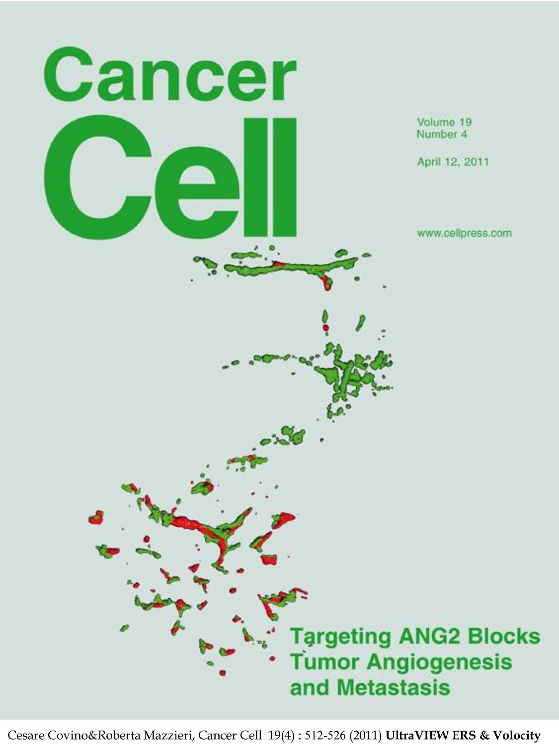
Volocity
Probably the most powerful and user-friendly software designed to provide you with rapid, interactive, high resolution volume rendering of your 3D and 4D data sets. Volocity is installed in a dedicated Windows Xp 64bit off-line workstation.

Huygens software
High-quality programs intended for restoration, interactive analysis and volume visualization of 2D, 3D, multi-channel and time series images from fluorescence microscopes. Huygens Software is also installed in a dedicated Windows Xp 64bit off-line workstation.

Softworx
SoftWorx is acquisition/analysis software of DeltaVision RT Deconvolution System installed in a dedicated off-line workstation, Linux RedHat-based. SoftWorx provides data management, visualization and image restoration and deconvolution (with measured PSF) of DeltaVision RT datasets.

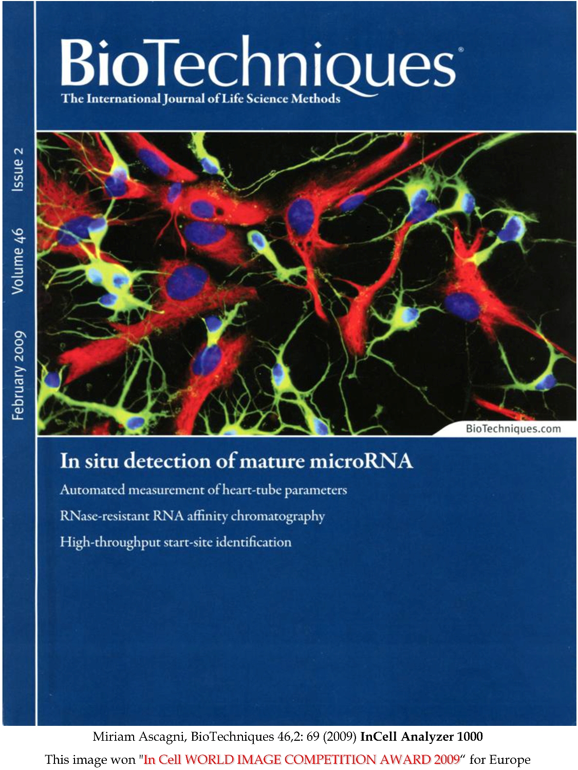
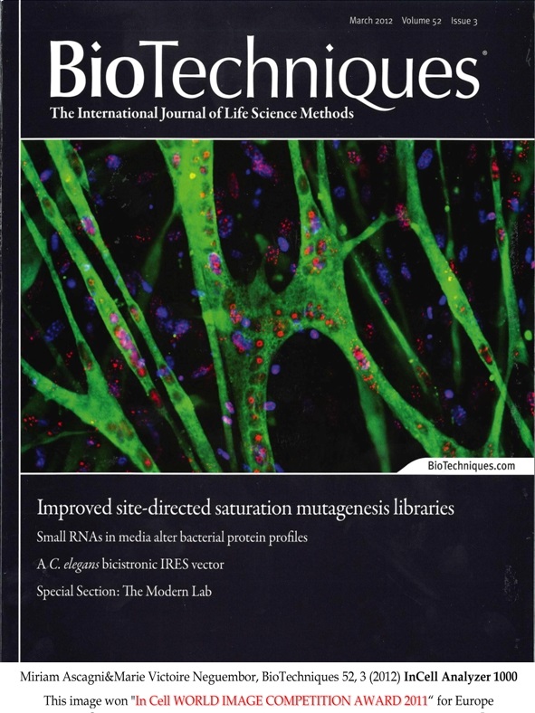
IN Cell Investigator
Automated cell imaging devices, such as the IN Cell Analyzer 1000, generate high volumes of data. IN Cell Investigator software was designed to efficiently analyze and interpret those amounts of data from live and fixed cell assays. IN Cell Investigator Software can work with images recorded by other instrumentations (.tiff format) when using the IN Cell Translator Software.

Oko-Vision
Oko-Vision software (from OkoLab) is a modular software specifically designed for live cells multidimensional experiments. This software easily allows you to select the images of the sequence that has to be analyzed, to plot and graph data sets and perform data aggregation and statistical analysis, especially for Cell Tracking and Wound Healing assay experiments.

Arivis
Arivis 4D (from Arivis) is a powerful software solution for visualization, sharing, analysis and presentation of multi-channel and multi-dimensional (2D, 3D and 4D) image data of almost unlimited size, independently of available RAM. Arivis 4D can easily import most image formats from microscopes and allows high performance interactive 3D / 4D rendering visualization and data processing with annotation and analysis with powerful pipelines. It could also integrate custom custom workflows via Matlab API or Python scripting.
Check out our Freeware BioImaging Software Web-Case
Amazing software and image analysis tools often have one big fault...they are very very expensive! Not always (unfortunately), but often (fortunately) scientists, researchers, programmers or software developers build and allow free use of very interesting and useful software or imaging tools. This page is a free space where you can point out your free software for the whole community. If you have any free software to indicate please send an email to Cesare Covino and we will be glad to publish it on our Freeware BioImaging Software Web-Case page (Last update: May 27th, 2009).